
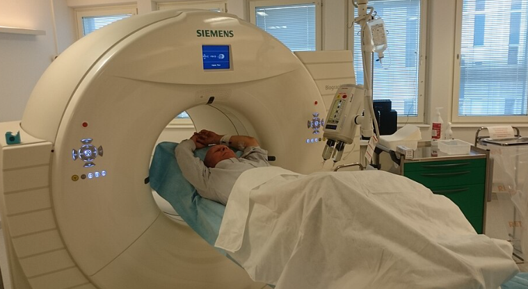
Researchers at the Medical University of Vienna found that PSMA-PET/CT imaging could help doctors identify aggressive prostate cancer and potentially reduce unnecessary biopsies.
A specialized imaging scan detected 91% of clinically significant prostate cancers in a study of 194 patients, offering a possible new way to improve early diagnosis. Researchers at the Medical University of Vienna found that the technique could help doctors determine which patients need a prostate biopsy and which might safely avoid the procedure.
The findings were published August 25, 2026, in The Lancet Medical Imaging & Theranostics. The research team, led by nuclear medicine specialist Marcus Hacker, examined how effectively a scan called PSMA-PET/CT could identify prostate cancer in men already suspected of having the disease.
Of 77 patients with clinically significant prostate cancer confirmed through tissue examinations, the scan correctly identified 70. That represents a detection rate of approximately 91%, although seven patients with significant cancer were not identified by the imaging method.
The results also showed that a normal scan was a potentially useful sign. Among patients whose scans did not indicate clinically significant cancer, 91% were found not to have such tumors.
Researchers believe these findings could eventually help doctors make better decisions about performing biopsies. However, the test is not yet established as a replacement for traditional diagnostic procedures, and further studies are necessary before its routine use for this purpose.
How the new imaging approach works
The imaging technique combines positron emission tomography, known as PET, with computed tomography, or CT. Together, these methods allow doctors to examine suspicious tissue and determine where it is located inside the body.
During the procedure, patients receive a small amount of a radioactive imaging substance. The substance attaches to prostate-specific membrane antigen, or PSMA, a protein found in particularly high amounts on many prostate cancer cells.
The PET component detects the radioactive signal, helping specialists identify areas that may contain cancer. The CT component provides detailed images of surrounding structures so doctors can determine the location of suspicious findings.
This combined approach gives physicians information about both possible cancer activity and the anatomy of the prostate. It may be particularly helpful when standard tests produce uncertain results.
PSMA-PET/CT is not an entirely new technology. It has already been used in prostate cancer care, particularly to investigate cancer that has spread or returned after treatment.
What makes the Vienna research important is its focus on patients undergoing their initial evaluation for suspected prostate cancer. The researchers examined whether the same imaging technology could provide useful information before a biopsy.
Why researchers want to improve prostate cancer diagnosis
Prostate cancer is commonly investigated after a blood test reveals an elevated level of prostate-specific antigen, or PSA. Although a high PSA level can be associated with cancer, it can also occur because of other prostate conditions.
An elevated result therefore does not automatically mean a patient has cancer. Doctors consider additional information, including medical history, physical examinations and imaging findings, before deciding whether a biopsy is appropriate.
Magnetic resonance imaging, or MRI, already plays an important role in evaluating many patients with suspected prostate cancer. Doctors often use MRI findings to identify suspicious areas and guide tissue sampling.
A biopsy allows specialists to examine prostate tissue under a microscope. It remains an important method for confirming cancer and determining how aggressive the disease appears.
However, biopsies are invasive and can cause bleeding, discomfort and infections. Some patients undergo the procedure only to learn that they do not have clinically significant cancer.
The Vienna study suggests that PSMA-PET/CT could eventually provide another source of information when physicians assess whether tissue sampling is necessary.
Scan also identified patients with aggressive disease
Researchers also examined what happened to participants after their initial scans. The median follow-up period was 35 months, meaning half the patients with follow-up data were observed for longer and half for shorter periods.
Among 176 patients included in this part of the analysis, 24 met the study’s criteria for an aggressive course of prostate cancer. The original scans had already identified 23 of those patients, representing approximately 96%.
This finding suggests that the imaging technique may help identify patients who are at greater risk of serious disease. However, it does not mean that the scan predicted 23 entirely new tumors that developed years later.
The researchers defined an aggressive disease course using several medical outcomes. These included metastatic cancer at the initial evaluation, a rise in PSA after prostate removal and certain treatment decisions involving hormonal therapy or chemotherapy.
The distinction matters because researchers were evaluating whether the initial scan was associated with later clinical outcomes. They were not demonstrating that the technology could detect cancers before those cancers existed.
The study also found that PSMA-PET/CT detected 84 of 106 prostate cancers when all confirmed cancers were considered, including less aggressive cases. That represents an overall sensitivity of approximately 79%.
The higher 91% detection rate applied specifically to clinically significant prostate cancer. These are tumors considered more likely to require medical attention than low-grade cancers.
A normal scan may help avoid some biopsies
One of the study’s most important findings involved the accuracy of negative scan results. Researchers reported a 91% negative predictive value for clinically significant prostate cancer.
In practical terms, this means that 91% of patients with a negative result did not have clinically significant cancer in the study population. It also means a normal scan could not completely rule out the disease.
The scan’s specificity was approximately 57% for clinically significant cancer. Specificity measures how often a test correctly identifies patients who do not have the condition.
That result shows that the technology also produced positive findings in some patients who did not have clinically significant disease. A suspicious scan therefore cannot be treated as definitive proof that a dangerous tumor is present.
The positive predictive value was 58%, meaning that slightly more than half of patients with positive scans had clinically significant cancer under the study’s criteria.
These numbers are important when considering whether the technology could reduce biopsies. A useful screening or diagnostic tool must balance the risk of missing serious disease against the risk of unnecessary procedures.
Study leader Marcus Hacker said the results indicate that PSMA-PET/CT may provide valuable information early in the diagnostic process. He emphasized that the reliability of a normal scan could help physicians decide which patients genuinely need a biopsy.
Further research is needed
The research was a secondary analysis of the RAPID clinical trial, a randomized phase 3 study conducted at Vienna General Hospital. Investigators analyzed imaging results collected from participants evaluated for suspected prostate cancer.
The analysis involved 194 patients at a single medical center. Although the original clinical trial was randomized, this particular assessment of PSMA-PET/CT results was conducted afterward.
That distinction limits how confidently the findings can be applied to broader patient populations. Larger studies involving multiple hospitals and more diverse participants will help determine whether similar results can be achieved consistently.
Researchers must also evaluate the practical costs of using PSMA-PET/CT more frequently during initial prostate cancer investigations. PET imaging requires specialized equipment and radioactive tracers that may not be available at every medical facility.
Another consideration is how the scan would fit alongside existing tests, including PSA measurements and MRI. The research does not establish that PSMA-PET/CT should replace these methods.
A separate 2026 clinical trial published in The Lancet Oncology, called PRIMARY2, also examined PSMA-PET/CT in men with suspected prostate cancer and uncertain or nonsuspicious MRI results.
In that Australian study, 163 of 331 patients assigned to the imaging-based approach avoided a biopsy. Researchers found that the strategy did not perform worse than standard biopsy for detecting clinically significant prostate cancer within the trial’s specified statistical limits.
Those findings provide additional evidence that PSMA-based imaging could help refine diagnostic decisions. However, the two studies examined different patient groups and used different research methods, so their results should not be considered interchangeable.
For patients with elevated PSA levels, the Vienna findings represent a possible improvement in how doctors investigate suspicious results. They do not establish that a negative scan makes further medical evaluation unnecessary.
The research team believes PSMA-PET/CT could eventually become part of a more targeted diagnostic process. Whether it can safely reduce biopsies in everyday medical practice will depend on additional clinical trials and evidence about its accuracy, cost and availability.
IMAGE: Source Own work Author Då.nu CC4